
Each foot has two peroneal tendons that help stabilize the foot and ankle. One peroneal tendon attaches to the outside of your midfoot, while the other runs under the foot and attaches to the inside of the arch. These tendons help stabilize the foot and prevent sprains and strains, but like any other tendon, they can become inflamed from overuse.
In today’s post, we look closely at how peroneal tendonitis develops and how you can diagnose and treat it.
Anatomy of the peroneal tendon
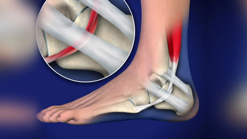
Two peroneal muscles, Peroneus longus, and Peroneous brevious are located in the outer part of the legs.
These two muscles’ tendons pass below the ankle’s back outside the ankle. The passage of these tendons behind the outer ankle is through a bony depression and groove, and a wide band of tissue covers this groove.
In this way, a tunnel is formed, the bottom of which is bone, and the top is made of a strong band of tissue called the Retinaculum. The contraction of the peroneal muscles causes their tendons to rise, and then the ankle goes down and rotates outward.
Sometimes this tendon or any tendons in your foot may cause some serious problems. In such cases, you’ll need to visit a doctor. There are so many good doctors in Dubai. Also, if you are searching for treatment for toe fractures, you can find the best one.
✔️ Read More: What is ankle disc osteochondritis? How to treat it?
What is the peroneal tendon?
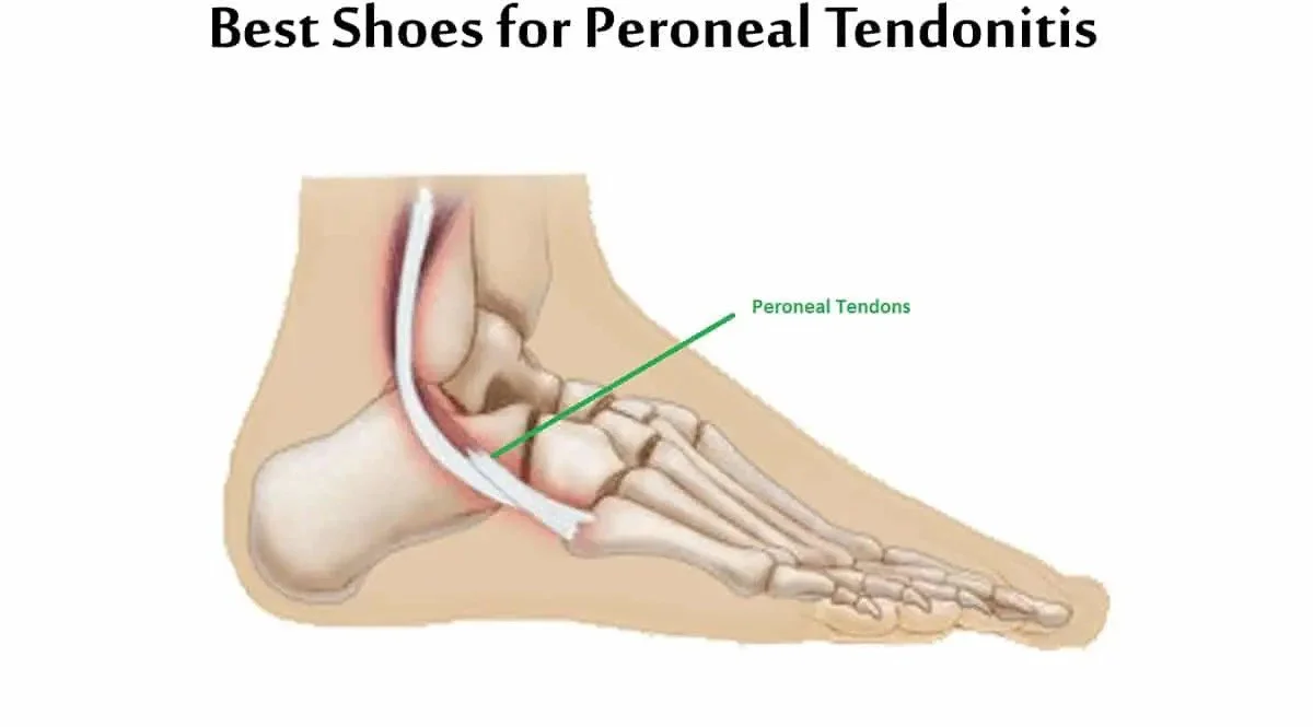
The peroneal tendons are the tendons that connect the muscles of the outer part of the calf muscle to the foot. Two large peroneal muscles (peroneus brevis and peroneus longus) are located outside the leg, right next to the muscles behind the leg. The muscles are attached to the bone by tendons, which, of course, run along the outside of the ankle and attach to the foot.
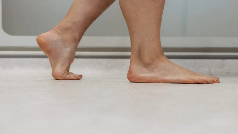
The peroneal muscles play an important role in turning the sole upwards. That is the movement of shaking the foot to the outside of the ankle. In normal walking, the movement of the peroneal muscles is balanced by the muscles that invert the foot (moving the foot inward from the ankle). Two peroneal tendons are very closely related.
In fact, one of them is on top of the other just behind the fibula. This close relationship is to contribute to some problems with the peroneal tendons, as they rub against each other on the back of the ankle. Peroneal tendons sometimes face injuries, or you might face peroneal tendonitis. In such cases, you should go for treatment of peroneal tendon injuries in Dubai.
What are the symptoms of peroneal tendonitis?
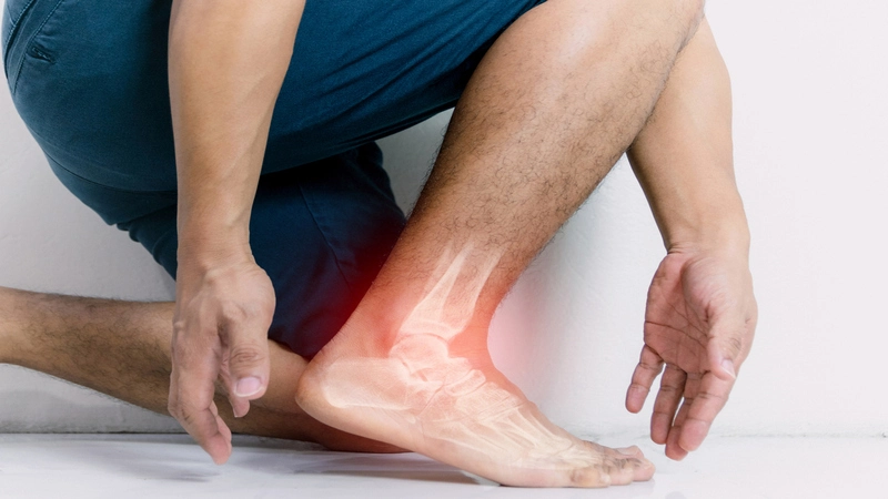
Peroneal tendon injuries can be acute (with a sudden event) or chronic (over time). These injuries happen mostly to people who participate in sports that involve frequent ankle movements.
In addition, people whose foot arch is higher than normal are more at risk of peroneal tendon injuries. The main types of peroneal tendon injuries are tendonitis (inflammation of the tendon), rupture, and tendon instability.
1- Tendonitis
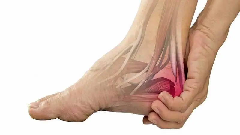
It is inflammation of one of the tendons or both of them. Inflammation is caused by activities that involve repetitive use of the tendon, overuse of the tendon, and injury (such as a sprained ankle). Symptoms of tendonitis include:
- Pain
- Swelling
- Feeling warm when touched
2- Peroneal tendon rupture
Severe tears are caused by repetitive activity or injury. Immediate symptoms of these severe tears include:
- Pain
- Inflation
- Weakness or instability of the foot and ankle
Over time, these tears can lead to a change in the shape of the foot where the angle of the arch of the foot increases (the sole becomes more concave).
Chronic tendon injury (tendinosis) usually occurs due to overuse and over long periods (usually several years).
In chronic tears, the tendon is like a rubber band that is stretched until it narrows and finally tears. Suffering from an excessive angle of the foot arch also puts a person at risk of suffering from chronic tendon rupture. Symptoms of chronic tendon tears include the following:
- Single pain (occasionally) in the outer part of the ankle
- Weakness or instability in the ankle
- Increasing the angle of the foot arch
3- Dislocation of the foot tendon
A sprain means that one or both of the tendons are out of their normal position. In some cases, sprains are caused by a congenital condition where a person is born with a bone or muscle shape change. In other cases, sprains, such as a sprained ankle, occur after an injury.
Injury or damage to the tissue (retinaculum) that stabilizes tendons can lead to chronic tendon rupture. Dislocation symptoms include the following:
- The sensation of tendon vibration in the areas of the ankle bone
- Occasional pain in the back of the ankle bone.
- Ankle weakness or instability
Early sprain treatment is critical because a dislocated tendon is more prone to tearing. So if you feel this slipping sensation in your foot, consult a foot surgeon in Dubai immediately.
✔️ Read More: What is an ankle sprain? Examining its treatment methods
Peroneal tendon injury diagnosis
An initial evaluation by a foot and ankle surgeon is recommended because peroneal tendon injuries are sometimes misdiagnosed, and a person’s condition may worsen without treatment.
To diagnose a peroneal tendon injury, the surgeon will examine the foot and look for pain, instability, swelling, heat, and weakness outside the ankle. In addition, radiography or other advanced imaging methods may be needed to assess the injury fully.
The foot and ankle surgeon will also look for signs of ankle sprains and other related injuries that are sometimes associated with peroneal tendon injuries. Getting a proper diagnosis is important because persistent ankle pain and discomfort after a simple sprain can indicate other problems.
Don’t forget to visit a reputable doctor. There are so many good doctors in Dubai for treating different injuries related to the feet, especially the treatment of sesamoid injuries.
Causes of peroneal tendon injury
Tendons are attached to muscles, and a tendon is at the end of each muscle. The other end of the tendon connects to the bone. So muscles are connected to bones through tendons.
The ankle sprain is the main cause of partial dislocation of the peroneal tendon. During an ankle sprain, the foot usually rotates inward, and this severe rotation causes injury and possibly rupture of the ligaments around the ankle. It also causes stretching of the peroneal tendon and damage to the retinaculum of the ankle.
With damage to the retinaculum, the tendon comes out of its groove, but of course, it returns to its place. It is called tendon dislocation if the tendon cannot return to its original position.
During the ankle’s initial sprain, the ligament damage’s intensity may be so high that the damage to the retinaculum is not noticed. Still, when the sprain symptoms subside, partial dislocation symptoms appear after a while.
Peroneal tendon sprains are rare and occur mostly during sports in which the foot deviates strongly upward and inward, such as skiing, ice skating, and soccer.
During the ankle’s strong upward and inward movement, the peroneal muscles are strongly contracted to prevent further deviation of the foot. This strong contraction causes the pressure of the peroneal tendons on the retinaculum and, consequently, tears. As the retinaculum is torn, the tendon dislocates.
In some people, there are changes in the ankle that make the person prone to partial peroneal dislocation. Sometimes the depth of the bony groove where the peroneal tendon passes behind the external ankle is small. Sometimes, the retinaculum’s tissue band is loose, making the tendon tend to move and leave the groove more.
✔️ Read More: What is the Avascular Necrosis of the Talus?
Treatment of peroneal tendonitis
The general principle for treating foot and ankle tendonitis is to rest the injured area so the body can heal. This usually takes weeks to months.
The best treatment methods for tendinitis or peroneal tendon inflammation include the following. Consult your doctor before starting treatment:
Medicine
The main goals of drug treatment are to relieve pain, restore mechanics, and return the patient to the level of participation in desired activities. Patients with peritoneal tendonitis can be treated with nonsteroidal anti-inflammatory drugs (NSAIDs) and reduced activity levels to relieve pain. There is insufficient evidence on the effectiveness of corticosteroids. Also, lateral heel wedges can help manage mild cases of peroneal tendonitis.
Management and treatment with physiotherapy
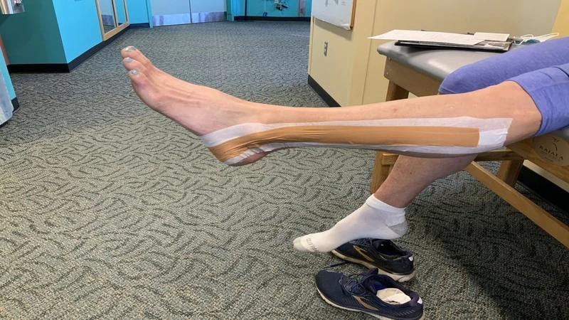
The treatment of peroneal tendon inflammation includes a program of stretching, strengthening, and mobilization (manual movement). Also, it includes manual therapy, strengthening and balancing exercises, using ice, and taping during contact sports.
If symptoms are severe, plastering of the area and immobilization with a ROM boot for ten days are prescribed. After the symptoms have resolved, the patient begins a rehabilitation program with a gradual increase in activity to reach their full level of activity. Other recommended treatments in physiotherapy are as follows:
- The use of a biomechanical ankle platform (BAPS)
- Deep tissue friction massage
- Ultrasound electrical stimulation
- Shock wave therapy (ESWT)
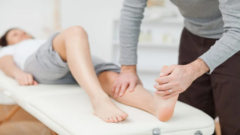
There is some evidence for manual therapy, especially the lateral movement of the hand on the calcaneus: to move the left calcaneus, the patient is placed on the left side while the calcaneus hangs off the table. The foot is kept in a neutral position, the ankle bone talus is kept fixed, and the therapist stretches from the middle to the sides (in a transverse position).
Stretching movements
You can start these exercises when you can stand on your injured leg with the heel on the floor, and you can equally put your full weight on both legs.
Stretching with a towel

Sit in a firm place and stretch your injured leg straight in front of you. Wrap a towel around your toes and the ball of your foot. Then pull the towel towards your body, keeping your leg straight. Hold this position for 15 to 30 seconds and then rest. Repeat this exercise three times.
Stretching the back of the calf in a standing position

Stand in front of the wall and place your hands on the wall. Hold your injured leg back while the heel is completely on the floor. Keep the other leg forward with the knee bent. Turn your back foot slightly inward (as if your toes are curled inward).
Lean gently against the wall until you feel a stretch in the back of your leg. Hold the stretch for 15 to 30 seconds. Return to the starting position. Repeat the movement three times. Do this exercise several times a day.
Stretching the soleus muscle or soleus muscle while standing
Stand against a wall and place your hands on the wall at chest height. The injured leg is moved back while the heel is kept on the ground. Keep the other leg forward with the knee bent. Turn your back foot slightly inward (as if your toe is turned inward).
Bend your back knee slightly and gently lean against the wall until you feel a stretch in the back of your injured leg. Hold the stretch for 15 to 30 seconds. Return to the starting position. Repeat the movement three times.
Achilles tendon strain
Stand on your toes (mostly on the ball under your toes) on a step. Pull your heels down toward the back step until you feel a stretch in the arch of your foot. Hold this position for 15 to 30 seconds and then rest. Repeat this movement three times.
✔️ Read More: Ankle instability: everything you need to know about it!
Surgery for peroneal tendon rupture
Peroneal tendon tears are uncommon and almost always occur in the peroneus brevis tendon. These tears are thought to result from two tendons’ problems. It is a blood supply issue. A peroneus brevis tear almost always occurs in a fluid-filled area where there is blood supply and, therefore, less nutrition to the tendon. Another issue is the tight connection between the peroneus brevis tendon and the peroneus longus tendon, which causes the peroneus brevis to get stuck between the peroneus longus tendon and the bone.
Many doctors try to treat peroneal tendon tears with the same treatments listed above for tendinitis. Unfortunately, many of these patients do not find lasting improvement in symptoms so surgery may be necessary. There are two surgical methods for peroneal tendon rupture:
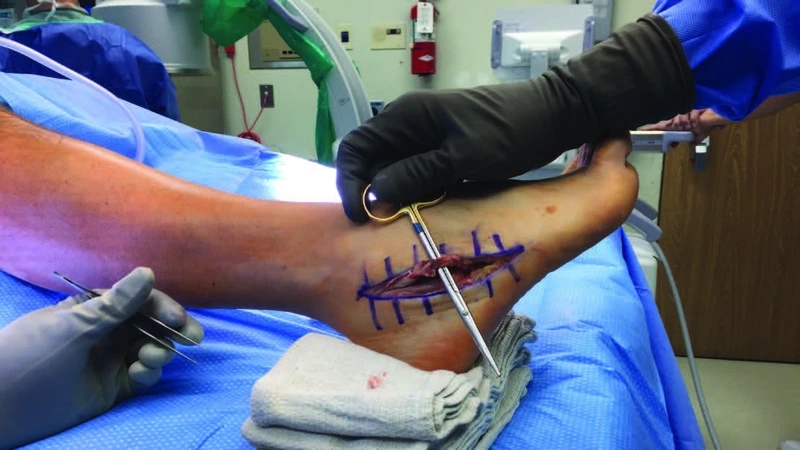
Tendon repair and debridement: The damaged tendon and the inflammatory tissue around it are removed during tendon debridement. A tendon tear can be repaired, and the tendon will “flatten” and regain its normal shape. Tendon debridement and repair is effective when less than 50% of the tendon is torn.
Tendon suture: A tenodesis is a procedure in which the damaged tendon is sutured to the normal tendon. In this case, the damaged part of the peroneus brevis (usually a few centimeters) is removed, and the left end is sutured to the adjacent peroneus longus tendon. This procedure is recommended for tears involving more than 50% of the tendon.
Postoperative recovery, depending on the type of surgery, includes several weeks of immobility and limited weight bearing. After a period of rest and immobility, physical therapy can be started. Depending on the extent and severity of the surgery, the total recovery time is usually 6 to 12 weeks. Risks of surgery include infection, dryness, and persistent pain. According to statistics, this surgery is very successful, with patients reporting a success rate of 85-95%.


